touchCARDIO touchCARDIO
Arrhythmia, Electrophysiology
Read Time: 2 mins

40/Electro-architectural differences between septal and non-septal post-infarct ventricular scars identified using ripple mapping

Copy Link
Published Online: Oct 9th 2012 European Journal of Arrhythmia & Electrophysiology. 2022;8(Suppl. 1):abstr40
Authors: P Calvert (Presenting Author) – Liverpool Heart & Chest Hospital, Liverpool; D Khanra – Liverpool Heart & Chest Hospital, Liverpool; S Hughes – Liverpool Heart & Chest Hospital, Liverpool; J Waktare – Liverpool Heart & Chest Hospital, Liverpool; S Modi – Liverpool Heart & Chest Hospital, Liverpool; M Hall – Liverpool Heart & Chest Hospital, Liverpool; D Todd – Liverpool Heart & Chest Hospital, Liverpool; S Mahida – Liverpool Heart & Chest Hospital, Liverpool; D Gupta – Liverpool Heart & Chest Hospital, Liverpool; V Luther – Liverpool Heart & Chest Hospital, Liverpool
Quick Links:
Article
Article Information
Article:

Background: Post-infarct septal scars are unique in their potential association with the native conduction system, but the influence of this relationship during ventricular tachycardia (VT) ablation has yet to be studied. High-density 3D mapping has offered new insights into the post-infarct ventricular electro-architecture. CARTO Ripple Maps (Biosense Webster) display myocardial activation as moving bars that can be superimposed on a 3D bipolar voltage map. We present a method using Ripple Mapping for delineating true non-conducting scar from low-voltage conducting borderzone tissue to describe observed electro-architectural differences between septal and non-septal scars.

Methods: Post-infarct VT ablations undertaken using Ripple Mapping in 12 consecutive patients (LVEF 31 ± 7%; median 5226 points) were studied. Substrate maps were collected with a Pentaray catheter with colour threshold between 3 and 5 in areas of low voltage <0.5 mV. We retrospectively analysed these cases and determined true non-conducting scar by sequentially reducing the voltage cut-off until no Ripple activation was seen within low-voltage areas. Regions with Ripple activation below the traditional 0.5 mV threshold were defined as conducting borderzone. We calculated the area of borderzone and scar in each case. We also calculated borderzone conduction speed by sampling two points of Ripple activation and utilising the time-caliper function to calculate distance over time. VT cycle lengths were also recorded. We then compared these characteristics between septal vs non-septal scars.

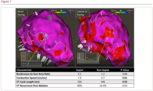
Results: Left ventricular (LV) scar was identified in all patients and was septal in 5 and non-septal in 7. Conducting borderzone tissue appeared more prevalent in septal vs non-septal scars (median borderzone-to-scar area ratio 3.2 vs 1.2; p=0.06). Borderzone conduction speed during atrial pacing was faster in septal scars (mean 7.4 cm/s vs 3.7 cm/s; p=0.01) and septal VTs were faster than non-septal VTs (mean cycle length 256 ms vs 346 ms; p=0.02). At 6-month follow-up, VT recurrence was more common in septal vs non-septal scar, but not to statistical significance, possibly due to low patient numbers (40.0% vs 14.3%; p=0.52). Figure 1 illustrates a septal scar from a patient with prior septal infarct and rapid VTs (cycle length 230 ms). Left: The LV substrate was mapped during atrial pacing. A white design line outlines the boundary of tissue <0.5 mV. Borderzone conducting tissue using Ripple Mapping was observed down to a threshold of 0.25 mV. Septal scar collocated within the vicinity of the native conduction system and appeared patchy and heterogeneous on the bipolar voltage map. A yellow circle highlights high-voltage activation ‘breakout’ possibly from a limb of surviving conduction tract. Right: A remap pacing from the basal lateral LV demonstrated lower voltages along the septum (yellow circle) perhaps as the conduction system is bypassed.

Conclusion: Septal scars have unique electro-architectural appearances on bipolar voltage mapping, likely due to their proximity to the nearby native conduction system. Potential interaction with surviving limbs of the conduction system may explain the observed increased borderzone conduction speed and shorter VT cycle lengths. This may result in potentially more challenging ablation.

Further Resources

Share this Article
Related Content In Electrophysiology
  • Copied to clipboard!
    accredited arrow-down-editablearrow-downarrow_leftarrow-right-bluearrow-right-dark-bluearrow-right-greenarrow-right-greyarrow-right-orangearrow-right-whitearrow-right-bluearrow-up-orangeavatarcalendarchevron-down consultant-pathologist-nurseconsultant-pathologistcrosscrossdownloademailexclaimationfeedbackfiltergraph-arrowinterviewslinkmdt_iconmenumore_dots nurse-consultantpadlock patient-advocate-pathologistpatient-consultantpatientperson pharmacist-nurseplay_buttonplay-colour-tmcplay-colourAsset 1podcastprinter scenerysearch share single-doctor social_facebooksocial_googleplussocial_instagramsocial_linkedin_altsocial_linkedin_altsocial_pinterestlogo-twitter-glyph-32social_youtubeshape-star (1)tick-bluetick-orangetick-red tick-whiteticktimetranscriptup-arrowwebinar Sponsored Department Location NEW TMM Corporate Services Icons-07NEW TMM Corporate Services Icons-08NEW TMM Corporate Services Icons-09NEW TMM Corporate Services Icons-10NEW TMM Corporate Services Icons-11NEW TMM Corporate Services Icons-12Salary £ TMM-Corp-Site-Icons-01TMM-Corp-Site-Icons-02TMM-Corp-Site-Icons-03TMM-Corp-Site-Icons-04TMM-Corp-Site-Icons-05TMM-Corp-Site-Icons-06TMM-Corp-Site-Icons-07TMM-Corp-Site-Icons-08TMM-Corp-Site-Icons-09TMM-Corp-Site-Icons-10TMM-Corp-Site-Icons-11TMM-Corp-Site-Icons-12TMM-Corp-Site-Icons-13TMM-Corp-Site-Icons-14TMM-Corp-Site-Icons-15TMM-Corp-Site-Icons-16TMM-Corp-Site-Icons-17TMM-Corp-Site-Icons-18TMM-Corp-Site-Icons-19TMM-Corp-Site-Icons-20TMM-Corp-Site-Icons-21TMM-Corp-Site-Icons-22TMM-Corp-Site-Icons-23TMM-Corp-Site-Icons-24TMM-Corp-Site-Icons-25TMM-Corp-Site-Icons-26TMM-Corp-Site-Icons-27TMM-Corp-Site-Icons-28TMM-Corp-Site-Icons-29TMM-Corp-Site-Icons-30TMM-Corp-Site-Icons-31TMM-Corp-Site-Icons-32TMM-Corp-Site-Icons-33TMM-Corp-Site-Icons-34TMM-Corp-Site-Icons-35TMM-Corp-Site-Icons-36TMM-Corp-Site-Icons-37TMM-Corp-Site-Icons-38TMM-Corp-Site-Icons-39TMM-Corp-Site-Icons-40TMM-Corp-Site-Icons-41TMM-Corp-Site-Icons-42TMM-Corp-Site-Icons-43TMM-Corp-Site-Icons-44TMM-Corp-Site-Icons-45TMM-Corp-Site-Icons-46TMM-Corp-Site-Icons-47TMM-Corp-Site-Icons-48TMM-Corp-Site-Icons-49TMM-Corp-Site-Icons-50TMM-Corp-Site-Icons-51TMM-Corp-Site-Icons-52TMM-Corp-Site-Icons-53TMM-Corp-Site-Icons-54TMM-Corp-Site-Icons-55TMM-Corp-Site-Icons-56TMM-Corp-Site-Icons-57TMM-Corp-Site-Icons-58TMM-Corp-Site-Icons-59TMM-Corp-Site-Icons-60TMM-Corp-Site-Icons-61TMM-Corp-Site-Icons-62TMM-Corp-Site-Icons-63TMM-Corp-Site-Icons-64TMM-Corp-Site-Icons-65TMM-Corp-Site-Icons-66TMM-Corp-Site-Icons-67TMM-Corp-Site-Icons-68TMM-Corp-Site-Icons-69TMM-Corp-Site-Icons-70TMM-Corp-Site-Icons-71TMM-Corp-Site-Icons-72