touchCARDIO touchCARDIO
Interventional Cardiology
Read Time: 4 mins

Evaluating Equivocal Left Main Stenosis with Fractional Flow Reserve – Not all Daughter Vessels are Created Equally

Copy Link
Published Online: Dec 9th 2019 Heart International. 2019;13(2):38–40 DOI: https://doi.org/10.17925/HI.2019.13.2.38
Authors: Ahmed Almomani, Abdul-rahman Abdel-karim, Barry F Uretsky
Quick Links:
Abstract
Article
Article Information
Abstract:
Overview

Fractional flow reserve (FFR) evaluation of intermediate left main coronary artery (LMCA) stenosis has been validated in clinical decision-making. However, the size of the daughter vessel, in which the FFR transducer is placed and the amount of myocardium it subtends, has received less attention. We present a case that demonstrates the importance of transducer location, size of the daughter vessel and the amount of subtended myocardium in evaluation of LMCA stenosis, pointing out potential pitfalls.

Keywords

Left main disease, fractional flow reserve, coronary revascularisation, intravascular ultrasound, angiography

Article:

Fractional flow reserve (FFR) has been a useful tool in guiding decision-making in intermediate left main coronary artery (LMCA) stenosis, particularly in the absence of downstream disease.1 Multiple caveats have been raised about the use of FFR with significant downstream disease and the interpretation of the significance of the LMCA stenosis.2 On the other hand, very little emphasis has been placed on the importance of the FFR transducer location and the amount of myocardium subtended, presumably because, in most cases, the left anterior descending (LAD) and left circumflex (Cx), both supply a large amount of myocardium that would easily clarify whether blood flow is limited from a LMCA stenosis. However, this may not always be true as illustrated by this case.

Case report

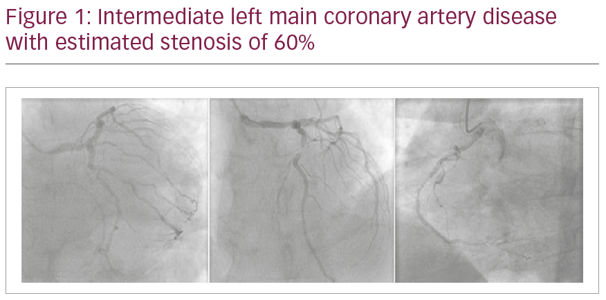
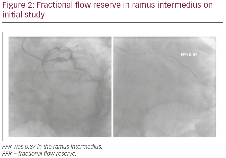
A 69-year-old man was referred for angiography for Canadian Cardiovascular Society (CCS) class III angina. He previously had stents implanted in his right coronary artery, LAD and first obtuse marginal branch. A coronary angiogram showed intermediate LMCA stenosis (Figure 1). Due to severe radial artery spasm and vessel tortuosity, catheter manipulation was challenging. As such, it was elected to perform FFR of the LMCA through the diagnostic catheter. The pressure wire could only be advanced into the ramus intermedius (RI) and pressure transducer for few millimetres distal to the lesion, and intravenous adenosine (140 mcg/kg/min) was administered to achieve haemodynamic response; FFR was 0.87 (Figure 2). On the basis of the unchanged coronary anatomy, compared to a previous angiogram 3 years earlier, except for mild angiographic progression of the LMCA stenosis and the non-ischaemic FFR value, it was elected to treat the patient medically.


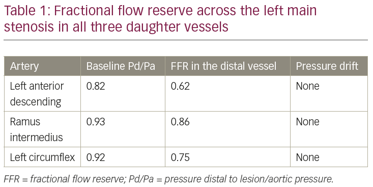
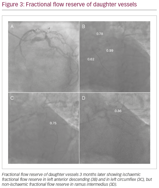
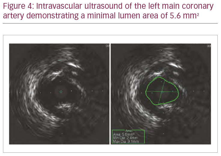
One month later, the patient reported improved but persistent CCS II angina. On the basis of the symptoms and angiographic severity of the left main lesion, the patient was brought back for FFR and intravascular ultrasound of the LMCA lesion through the femoral approach to overcome the technical difficulties faced in the previous procedure. FFR of the LMCA lesion was performed in all three daughter vessels (LAD, Cx and RI). Again, FFR was non-ischaemic (0.86) in the RI consistent with the previous assessment with the pressure wire greater than 10 mm distal to the lesion. However, FFR was in the ischaemic range for both the LAD and the Cx (Table 1, Figure 3). Intravascular ultrasound of LMCA showed a minimal luminal area of 5.6 mm2 (Figure 4). The patient was referred for coronary artery bypass surgery.


Discussion

FFR-guided percutaneous coronary intervention in non-left main lesions has shown better long-term clinical outcomes compared to angiography alone in stable ischaemic heart disease.3,4 Furthermore, numerous studies support the use of FFR to assess intermediate LMCA coronary stenosis.1,5–7 However, as with any diagnostic test, the technique and limitations of its applications should be understood.

The reliability of FFR for simple LMCA lesions is rarely an issue, but interpreting the LMCA FFR in the presence of significant downstream branch lesions, such as an LAD stenosis, is a recognised challenge. LMCA and the downstream lesions act as serial lesions, and the true flow across the LMCA is potentially reduced by a severe downstream stenosis, resulting in false elevation of the LMCA FFR when measured in the unobstructed vessel.2,8 Less emphasis has been placed on the importance of the location of the FFR transducer in the absence of downstream stenosis and its relation to the amount of myocardium supplied by the daughter branch.9 For an accurate FFR, maximal hyperaemia must be achieved across the LMCA stenosis. Flow through the LMCA is the sum of the flow in all daughter branches, the magnitude of flow being proportional to the size of each artery’s viable myocardial bed.

This case points out that the size of the daughter vessel and the amount of myocardium it subtends is a factor in determining the measured FFR. In this particular case, the RI was small with a small subtended myocardium, and despite the LMCA lesion, the blood flow required during maximal hyperaemia was adequate. In small coronary arteries pressure recovery occurs within 1–2 mm distal to stenosis,10 and the fact that the transducer could only be advanced few millimetres distal to the lesion should not have affected the FFR measurement. Additionally, the fact that the FFR value on the second study was essentially the same as the first is in keeping with the accuracy of the first measurement. On the other hand, both the Cx and LAD were larger vessels with larger subtended myocardium and both showed ischaemic FFR. Thus, it is important to keep in mind that the pressure transducer needs to be in a larger daughter vessel of the LMCA when measuring FFR in order to be used appropriately for clinical decision-making.

Conclusion

FFR is well accepted as a valuable tool in clinical decision-making of intermediate left main stenosis. This case illustrates an important consideration in its use. Specifically, if the pressure transducer is in a small daughter vessel, typically a small RI or Cx, the FFR value may be in the non-ischaemic range due to the limited area of myocardium subtended by the vessel when, in fact, FFR measured in a larger vessel may show ischaemia. The case emphasises the need to measure FFR in all the daughter vessels, especially those with the largest myocardial territory.

Article Information:
Disclosure

Ahmed Almomani, Abdul-rahman Abdel-karim and Barry Uretsky have no financial or non-financial relationships or activities to declare in relation to this article.

Compliance With Ethics

This study involves a review of the literature and did not involve any studies with human or animal subjects performed by any of the authors.

Review Process

Double-blind peer review.

Authorship

The named authors meet the International Committee of Medical Journal Editors (ICMJE) criteria for authorship of this manuscript, take responsibility for the integrity of the work as a whole, and have given final approval for the version to be published.

Correspondence

Barry F Uretsky, Central Arkansas Veterans Healthcare System, 4300 West Seventh Street, Little Rock, AR 72205, USA. E: buretsky@gmail.com

Support

No funding was received in the publication of this article.

Received

2019-04-03

References

  1. Hamilos M, Muller O, Cuisset T, et al. Long-term clinical outcome after fractional flow reserve-guided treatment in patients with angiographically equivocal left main coronary artery stenosis. Circulation. 2009;120:1505–12.
  2. Daniels DV, van’t Veer M, Pijls NH, et al. The impact of downstream coronary stenoses on fractional flow reserve assessment of intermediate left main disease. JACC Cardiovasc Interv. 2012;5:1021–5.
  3. De Bruyne B, Pijls NH, Kalesan B, et al. Fractional flow reserve-guided PCI versus medical therapy in stable coronary disease. N Engl J Med. 2012;367:991–1001.
  4. Pijls NH, Fearon WF, Tonino PA, et al. Fractional flow reserve versus angiography for guiding percutaneous coronary intervention in patients with multivessel coronary artery disease: 2-year follow-up of the FAME (fractional flow reserve versus angiography for multivessel evaluation) study. J Am Coll Cardiol. 2010;56:177–84.
  5. Bech GJ, Droste H, Pijls NH, et al. Value of fractional flow reserve in making decisions about bypass surgery for equivocal left main coronary artery disease. Heart. 2001;86:547–52.
  6. Jasti V, Ivan E, Yalamanchili V, et al. Correlations between fractional flow reserve and intravascular ultrasound in patients with an ambiguous left main coronary artery stenosis. Circulation. 2004;110:2831–6.
  7. Lindstaedt M, Yazar A, Germing A, et al. Clinical outcome in patients with intermediate or equivocal left main coronary artery disease after deferral of surgical revascularization on the basis of fractional flow reserve measurements. Am Heart J. 2006;152:156.e1–9.
  8. Fearon WF, Yong AS, Lenders G, et al. The impact of downstream coronary stenosis on fractional flow reserve assessment of intermediate left main coronary artery disease: human validation. JACC Cardiovasc Interv. 2015;8:398–403.
  9. Echavarría-Pinto M, van de Hoef TP, Nijjer S, et al. Influence of the amount of myocardium subtended to a coronary stenosis on the index of microcirculatory resistance. Implications for the invasive assessment of microcirculatory function in ischaemic heart disease. EuroIntervention. 2017;13:944–52.
  10. Ofili EO, Labovitz AJ, Kern MJ. Coronary flow velocity dynamics in normal and diseased arteries. Am J Cardiol. 1993;71:3–9D.

Further Resources

Share this Article
Related Content In Interventional Cardiology
  • Copied to clipboard!
    accredited arrow-down-editablearrow-downarrow_leftarrow-right-bluearrow-right-dark-bluearrow-right-greenarrow-right-greyarrow-right-orangearrow-right-whitearrow-right-bluearrow-up-orangeavatarcalendarchevron-down consultant-pathologist-nurseconsultant-pathologistcrosscrossdownloademailexclaimationfeedbackfiltergraph-arrowinterviewslinkmdt_iconmenumore_dots nurse-consultantpadlock patient-advocate-pathologistpatient-consultantpatientperson pharmacist-nurseplay_buttonplay-colour-tmcplay-colourAsset 1podcastprinter scenerysearch share single-doctor social_facebooksocial_googleplussocial_instagramsocial_linkedin_altsocial_linkedin_altsocial_pinterestlogo-twitter-glyph-32social_youtubeshape-star (1)tick-bluetick-orangetick-red tick-whiteticktimetranscriptup-arrowwebinar Sponsored Department Location NEW TMM Corporate Services Icons-07NEW TMM Corporate Services Icons-08NEW TMM Corporate Services Icons-09NEW TMM Corporate Services Icons-10NEW TMM Corporate Services Icons-11NEW TMM Corporate Services Icons-12Salary £ TMM-Corp-Site-Icons-01TMM-Corp-Site-Icons-02TMM-Corp-Site-Icons-03TMM-Corp-Site-Icons-04TMM-Corp-Site-Icons-05TMM-Corp-Site-Icons-06TMM-Corp-Site-Icons-07TMM-Corp-Site-Icons-08TMM-Corp-Site-Icons-09TMM-Corp-Site-Icons-10TMM-Corp-Site-Icons-11TMM-Corp-Site-Icons-12TMM-Corp-Site-Icons-13TMM-Corp-Site-Icons-14TMM-Corp-Site-Icons-15TMM-Corp-Site-Icons-16TMM-Corp-Site-Icons-17TMM-Corp-Site-Icons-18TMM-Corp-Site-Icons-19TMM-Corp-Site-Icons-20TMM-Corp-Site-Icons-21TMM-Corp-Site-Icons-22TMM-Corp-Site-Icons-23TMM-Corp-Site-Icons-24TMM-Corp-Site-Icons-25TMM-Corp-Site-Icons-26TMM-Corp-Site-Icons-27TMM-Corp-Site-Icons-28TMM-Corp-Site-Icons-29TMM-Corp-Site-Icons-30TMM-Corp-Site-Icons-31TMM-Corp-Site-Icons-32TMM-Corp-Site-Icons-33TMM-Corp-Site-Icons-34TMM-Corp-Site-Icons-35TMM-Corp-Site-Icons-36TMM-Corp-Site-Icons-37TMM-Corp-Site-Icons-38TMM-Corp-Site-Icons-39TMM-Corp-Site-Icons-40TMM-Corp-Site-Icons-41TMM-Corp-Site-Icons-42TMM-Corp-Site-Icons-43TMM-Corp-Site-Icons-44TMM-Corp-Site-Icons-45TMM-Corp-Site-Icons-46TMM-Corp-Site-Icons-47TMM-Corp-Site-Icons-48TMM-Corp-Site-Icons-49TMM-Corp-Site-Icons-50TMM-Corp-Site-Icons-51TMM-Corp-Site-Icons-52TMM-Corp-Site-Icons-53TMM-Corp-Site-Icons-54TMM-Corp-Site-Icons-55TMM-Corp-Site-Icons-56TMM-Corp-Site-Icons-57TMM-Corp-Site-Icons-58TMM-Corp-Site-Icons-59TMM-Corp-Site-Icons-60TMM-Corp-Site-Icons-61TMM-Corp-Site-Icons-62TMM-Corp-Site-Icons-63TMM-Corp-Site-Icons-64TMM-Corp-Site-Icons-65TMM-Corp-Site-Icons-66TMM-Corp-Site-Icons-67TMM-Corp-Site-Icons-68TMM-Corp-Site-Icons-69TMM-Corp-Site-Icons-70TMM-Corp-Site-Icons-71TMM-Corp-Site-Icons-72