touchCARDIO touchCARDIO
Arrhythmia
Read Time: 2 mins

23/The degree and spatial distribution of myocardial fibrosis can influence the underlying mechanism of ventricular fibrillation

Copy Link
Published Online: Oct 2nd 2008 European Journal of Arrhythmia & Electrophysiology. 2019;5(Suppl. 1):abstr23
Authors: BS Handa (Presenting Author) - Imperial College London, London, UK; X Li - Imperial College London, London, UK; CA Mansfield - Imperial College London, London, UK; RJ Jabbour - Imperial College London, London, UK; D Pitcher - Imperial College London, London, UK; A Chowdhury - Imperial College London, London, UK; NS Peters - Imperial College London, London, UK; FS Ng - Imperial College London, London, UK
Quick Links:
Article
Article Information
Article:

Background: Myocardial fibrosis is known to play a critical role in genesis and maintenance of ventricular fibrillation (VF). The quantity of fibrosis post myocardial infarction negatively correlates with survival. There is paucity of data on how the quantity and degree of fibrosis influences the mechanisms of VF. VF mechanisms remain debated, there are data supporting critical areas sustaining rotational drivers (RDs), while the contrary hypothesis is of disorganised myocardial activation only.

PurposeWe hypothesised that the underlying mechanism of VF is influenced by the degree and spatial distribution of myocardial fibrosis.
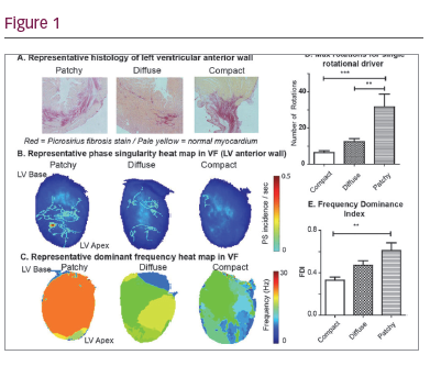
MethodsThirty-five Sprague-Dawley rats were allocated to undergo permanent left anterior descending (LAD) ligation (n=11), 20 mins LAD territory ischaemia-reperfusion (n=13) or in vivo angiotensin infusion (500ng/kg/min, n=11) to generate compact (CF), patchy (PF) and diffuse fibrosis (DF) models respectively. After a 4-week period of maturation, the hearts were explanted, Langendorff perfused and VF induced with burst pacing and 30μM pinacidil. Fibrillation dynamics were quantified using phase analysis, phase singularity (PS) tracking and our novel method of global fibrillation organisation quantification, frequency dominance index (FDI), which is defined as the power ratio of highest amplitude dominant frequency in the frequency spectrum.

ResultsVentricular fibrosis for each group was characterized and quantified (CF: 22.3 ± 3.2%, PF: 18.4 ± 4.2%, DF: 5.8 ± 1.3%, p = 0.046). VF was driven predominantly by disorganised activity in CF, PSs were detected 26 ± 7% of time comparative to 51.2 ± 4% in DF and 69.5 ± 8% in PF group (p = 0.001). PF stabilised RDs, average maximum rotations for a single RD in PF were 31.6 ± 7.1 comparative to 12.5 ± 1.7 in DF and 6.4 ± 1.1 in CF, p<0.001. The average maximum duration for a single RDs was significantly longer in PF (PF: 1231 ± 365 ms, DF: 568 ± 68 ms,
CF: 363 ± 41 ms, p = 0.014). Similarly, average rotations per RD were greater in the PF group (PF: 4.5 ± 0.7, DF: 3.3 ± 0.2, CF: 2.41 ± 0.3 rotations, p=0.013).Total number of RDs/second were much greater with PF (PF: 12.4 ± 2.0, DF: 5.4 ± 0.8, CF: 3.1 ± 1.1,p<0.001). Dominant frequency maps showed that in PF group VF was often driven by one or two well defined dominant frequencies, whereas in the DF and CF group multiple dominant frequencies were present. VF organisation measured by FDI was higher in PF
(PF: 0.61 ± 0.07, DF: 0.47 ± 0.04, CF: 0.33 ± 0.03, p = 0.004). RDs in DF showed a greater degree of meander comparative to PF (DF: 12.6 ± 1.4 versus PF: 9.3 ± 0.8 pixels, p = 0.024).

Conclusion: VF mechanisms occur along a spectrum between organised activity with discrete drivers and disorganized myocardial activation. The underlying VF mechanism can differ significantly dependent on the degree and pattern of fibrosis. Patchy fibrosis stabilises RDs with localization to discrete areas and sustains an organised form of VF comparative to CF where VF is largely disorganised. DF sustains an intermediate level of VF organisation. Characterising the degree and pattern of fibrosis in patient groups vulnerable to VF might be beneficial in identifying patients with targetable substrate.

Further Resources

Share this Article
Related Content In Arrhythmia
  • Copied to clipboard!
    accredited arrow-down-editablearrow-downarrow_leftarrow-right-bluearrow-right-dark-bluearrow-right-greenarrow-right-greyarrow-right-orangearrow-right-whitearrow-right-bluearrow-up-orangeavatarcalendarchevron-down consultant-pathologist-nurseconsultant-pathologistcrosscrossdownloademailexclaimationfeedbackfiltergraph-arrowinterviewslinkmdt_iconmenumore_dots nurse-consultantpadlock patient-advocate-pathologistpatient-consultantpatientperson pharmacist-nurseplay_buttonplay-colour-tmcplay-colourAsset 1podcastprinter scenerysearch share single-doctor social_facebooksocial_googleplussocial_instagramsocial_linkedin_altsocial_linkedin_altsocial_pinterestlogo-twitter-glyph-32social_youtubeshape-star (1)tick-bluetick-orangetick-red tick-whiteticktimetranscriptup-arrowwebinar Sponsored Department Location NEW TMM Corporate Services Icons-07NEW TMM Corporate Services Icons-08NEW TMM Corporate Services Icons-09NEW TMM Corporate Services Icons-10NEW TMM Corporate Services Icons-11NEW TMM Corporate Services Icons-12Salary £ TMM-Corp-Site-Icons-01TMM-Corp-Site-Icons-02TMM-Corp-Site-Icons-03TMM-Corp-Site-Icons-04TMM-Corp-Site-Icons-05TMM-Corp-Site-Icons-06TMM-Corp-Site-Icons-07TMM-Corp-Site-Icons-08TMM-Corp-Site-Icons-09TMM-Corp-Site-Icons-10TMM-Corp-Site-Icons-11TMM-Corp-Site-Icons-12TMM-Corp-Site-Icons-13TMM-Corp-Site-Icons-14TMM-Corp-Site-Icons-15TMM-Corp-Site-Icons-16TMM-Corp-Site-Icons-17TMM-Corp-Site-Icons-18TMM-Corp-Site-Icons-19TMM-Corp-Site-Icons-20TMM-Corp-Site-Icons-21TMM-Corp-Site-Icons-22TMM-Corp-Site-Icons-23TMM-Corp-Site-Icons-24TMM-Corp-Site-Icons-25TMM-Corp-Site-Icons-26TMM-Corp-Site-Icons-27TMM-Corp-Site-Icons-28TMM-Corp-Site-Icons-29TMM-Corp-Site-Icons-30TMM-Corp-Site-Icons-31TMM-Corp-Site-Icons-32TMM-Corp-Site-Icons-33TMM-Corp-Site-Icons-34TMM-Corp-Site-Icons-35TMM-Corp-Site-Icons-36TMM-Corp-Site-Icons-37TMM-Corp-Site-Icons-38TMM-Corp-Site-Icons-39TMM-Corp-Site-Icons-40TMM-Corp-Site-Icons-41TMM-Corp-Site-Icons-42TMM-Corp-Site-Icons-43TMM-Corp-Site-Icons-44TMM-Corp-Site-Icons-45TMM-Corp-Site-Icons-46TMM-Corp-Site-Icons-47TMM-Corp-Site-Icons-48TMM-Corp-Site-Icons-49TMM-Corp-Site-Icons-50TMM-Corp-Site-Icons-51TMM-Corp-Site-Icons-52TMM-Corp-Site-Icons-53TMM-Corp-Site-Icons-54TMM-Corp-Site-Icons-55TMM-Corp-Site-Icons-56TMM-Corp-Site-Icons-57TMM-Corp-Site-Icons-58TMM-Corp-Site-Icons-59TMM-Corp-Site-Icons-60TMM-Corp-Site-Icons-61TMM-Corp-Site-Icons-62TMM-Corp-Site-Icons-63TMM-Corp-Site-Icons-64TMM-Corp-Site-Icons-65TMM-Corp-Site-Icons-66TMM-Corp-Site-Icons-67TMM-Corp-Site-Icons-68TMM-Corp-Site-Icons-69TMM-Corp-Site-Icons-70TMM-Corp-Site-Icons-71TMM-Corp-Site-Icons-72