Despite improved treatment of atherosclerotic cardiovascular disease, coronary artery disease (CAD) remains the leading cause of death and disability worldwide.1 Treatment of traditional risk factors, mainly low-density lipoprotein (LDL) cholesterol, is the foundation of atherosclerotic risk reduction. However, a considerable residual risk for cardiovascular disease (CVD) still exists despite reducing LDL cholesterol.2 Beyond traditional risk factors, other drivers of residual risk have come to the forefront, including inflammatory, metabolic and pro-thrombotic pathways that contribute to recurrent cardiovascular events, and are often unrecognized in clinical practice.3
Coronary atherosclerosis is a multistep process starting with endothelial dysfunction and progressing to plaque formation, progression and rupture, which leads to thrombus formation and cardiovascular events.4 Eicosapentaenoic acid (EPA) has beneficial effects on many of these steps. In animal models, EPA suppressed the progression of atherosclerotic lesions and increased the cellular content of omega-3 polyunsaturated fatty acids (PUFAs) without increasing total cholesterol.5 This article reviews the clinical research using various cardiovascular imaging modalities on the effects of omega-3 fatty acids (OM3FAs) on atherosclerotic plaques.
Overview of omega-3 fatty acids, their indications and their biological effects
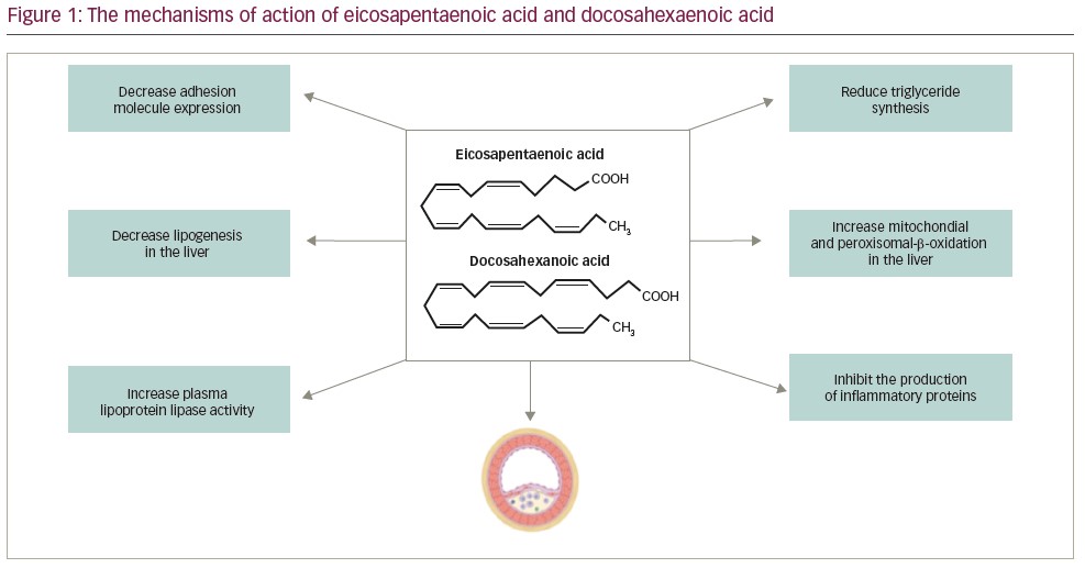
OM3FAs are unsaturated fatty acids with at least one double bond located between the third and fourth omega end carbon (Figure 1). There is emerging evidence from recent interventional clinical trials suggesting that omega-3 PUFAs – specifically EPA – confer cardiovascular protection.6,7 Currently, the three most clinically applicable OM3FAs are EPA, docosahexaenoic acid (DHA) and α-linolenic acid. These contain oils that originate from plant sources and can be found in fish, fish products, seeds, nuts, green leafy vegetables and beans.8 Currently, approved OM3FA formulations by US Food and Drug Administration include icosapent ethyl (IPE), omega-3-acid ethyl esters, omega-3 carboxylic acids and omega-3-acid ethyl esters type A.9–13 Omega-3 carboxylic acids and omega-3-acid ethyl esters type A have since been discontinued.13 Omega-3-acid ethyl esters, omega-3 carboxylic acids and omega-3-acid ethyl esters type A contain both EPA and DHA, whereas IPE contains ethyl esters of EPA only.14
Prescribed OM3FAs are approved for adults with very high triglyceride levels (≥500 mg/dL) as an adjunct to diet to decrease triglyceride levels and reduce the risk of cardiovascular events.15–17 IPE is approved as an adjunct to maximally tolerated statin therapy for reducing cardiovascular risk in patients with triglyceride levels >150 mg/dL and either established CVD or type 2 diabetes mellitus with ≥2 risk factors for CVD.18 In combination, it reduces risk more so than statins alone.19,20 OM3FA intake and/or supplementation not only reduces cardiovascular risk but is also beneficial in treating other conditions such as type 2 diabetes,20–22 cancer,23–25 Alzheimer’s disease and dementia,21,26 depression,21 heart failure,27 intervertebral disc degeneration,28 attention deficit hyperactivity disorder,29 menopausal night sweats,30 rheumatoid arthritis31 and non-alcoholic fatty liver disease.32
The mechanism of action of OM3FAs in lowering triglyceride levels is not fully understood; however, studies show that OM3FAs lower triglyceride levels by suppressing lipogenic gene expression, increasing beta-oxidation of fatty acids, increasing the expression of lipoprotein lipase, and influencing total body lipid accretion (Figure 1).33–35 Although some studies have reported comparable or even greater efficacy of DHA in reducing triglyceride levels and individual pro-inflammatory cytokines compared with EPA,36,37 a significant body of evidence suggests that EPA and DHA have markedly different effects on membrane structure, the inflammatory cascade, lipid oxidation, endothelial function and tissue distribution.36–38 Research indicates that DHA has a non-uniform effect on membrane fluidity and structure, resulting in cholesterol-rich regions that are pro-atherogenic and help form unstable atherosclerotic plaques.39
Furthermore, pure EPA (IPE) or high ratios of EPA are more effective in reducing oxidative stress than DHA.40 Pure EPA can lower triglyceride levels without raising LDL cholesterol levels, whereas DHA and EPA + DHA modestly increase LDL cholesterol levels in patients with elevated triglyceride levels, as seen in the ANCHOR and MARINE studies.41,42 This effect is hypothesized to be mediated by EPA enhancing the production of LDL receptors, which facilitates clearing LDL particles.43 Most notably, the anti-inflammatory action of EPA has been established in clinical trials, shown by significant reductions in serum inflammatory biomarkers that were not consistently seen in trials using EPA/DHA combinations.44,45 The distinct characteristics of EPA and DHA and their effect on atherogenesis, as evidenced by imaging trials, will be discussed further in this review.
Cardiovascular outcome trials of omega-3
fatty acids
In earlier studies, low doses of marine n-3 fatty acids (also known as OM3FA) did not reduce the incidence of major cardiovascular events.46–48 However, higher doses and the purified forms of EPA have shown promising results in reducing the risk of major adverse cardiovascular events (MACE) when used alongside statins in patients with elevated triglyceride levels, as seen in the REDUCE-IT (Reduction of cardiovascular events with icosapent ethyl-intervention) trial and JELIS (Japan EPA lipid intervention study).49,50
The VITAL (Vitamin D and omega-3 trial) trial, which emphasized primary prevention, was the largest randomized controlled trial investigating OM3FAs.48 With a median follow-up of 5.3 years, the VITAL study randomly assigned 25,000 patients to receive either 1 g/day of OM3FA, 2,000 IU of vitamin D3, both OM3FA and vitamin D3, or a dual placebo. When compared with placebo, OM3FA had no benefit in reducing MACE (myocardial infarction, stroke and cardiovascular death).48
Another randomized controlled trial, ASCEND (A study of cardiovascular events in diabetes), involved 15,480 participants with diabetes and no history of CVD.51 The effects of EPA, DHA and aspirin (separate part of the trial) on cardiovascular events were examined in this study. The subjects were randomly allocated to either 1 g/day of OM3FA or placebo (olive oil). ASCEND was also deemed a null omega-3 trial as the composite primary endpoint of serious vascular events (non-fatal myocardial infarction, non-fatal stroke, transient ischaemic attack and vascular death) was not significantly different in the omega-3 group compared with the placebo group after 7.4 years’ follow-up.51
Furthermore, the STRENGTH trial evaluated the impact of Epanova® (AstraZeneca, Wilmington, DE, USA; a fish oil containing EPA-DHA) in patients on optimal statin therapy with mixed dyslipidaemia and at high risk for CVD.52 This trial randomized 13,086 participants to receive 4 g of Epanova® daily or placebo (corn oil). This study found no significant reduction in the composite end point of cardiovascular death, myocardial infarction, stroke, coronary revascularization or hospitalization for unstable angina with Epanova®.52
Unlike the VITAL, ASCEND and STRENGTH trials, the REDUCE-IT trial used IPE, EPA in the ethyl ester form.49 The primary objective of the study was to determine whether IPE and statin combined were superior to statin therapy alone in preventing long-term cardiovascular events in high-risk patients with mixed dyslipidaemia. This study enrolled 8,179 participants with triglyceride levels 135–499 mg/dL, known CVD or diabetes and at least one other cardiovascular risk factor. Participants were randomly assigned to either 4 g/day IPE or placebo and followed for 5 years. The primary endpoint was a composite of cardiovascular death, non-fatal myocardial infarction, non-fatal stroke, coronary revascularization or unstable angina. IPE significantly reduced cardiovascular events by 25% compared with placebo.49
The use of different placebos in the STRENGTH and REDUCE-IT trials is cited as one of the reasons for these disparate results. In the STRENGTH study, corn oil was used as placebo, whereas mineral oil was used as placebo in REDUCE-IT. Mineral oil has potentially unfavourable cardiovascular effects and may have contributed to the increased event rate in the placebo group, hence enhancing the advantages of EPA. Additionally, mineral oil may interfere with statin absorption given the increase in lipids and C-reactive protein (CRP) levels seen in the control arm. However, it has to be noted that a risk reduction of 25% is too large to be attributable to a placebo. In the STRENGTH trial, corn oil was chosen as the placebo as it was deemed a neutral comparator with minimal impact on biochemical markers associated with cardiovascular risk. However, in the placebo arm there was a trend towards potential protection against atherosclerotic CVD due to lower LDL and CRP levels, which may have interfered with the final results of the study. The difference in the results of REDUCE-IT and STRENGTH is thought to arise from the biochemical differences between pure EPA and EPA + DHA, which is likely the main driver of cardiovascular benefit seen in these trials.49,52
The JELIS study looked at the effects of fish oil supplement EPA added to statin therapy in 18,645 patients with hypercholesterolaemia.50 In this open-label trial, participants were randomly assigned to receive either a statin alone or EPA (1,800 mg/day) plus statin. At a mean follow-up of 4.6 years, those who received EPA in addition to statin therapy saw an improvement in the primary composite outcome (fatal and non-fatal myocardial infarction, unstable angina, angioplasty, stenting, coronary artery bypass grafting or sudden cardiac death). The reduction in MACE seen with EPA in addition to statin therapy was independent of total cholesterol and LDL levels, which were reduced in both groups to a similar proportion.50
The REDUCE-IT and JELIS trials indicate that larger doses and pure EPA have distinct physiological effects compared with lower doses and EPA + DHA. In JELIS, participants with high (150 μg/L or more) treatment plasma EPA concentrations had a significantly lower risk of MACE than those with low (<87 μg/L) concentrations.53 Similarly, on-treatment higher plasma EPA levels were strongly linked with cardiovascular outcomes in the REDUCE-IT trial.54 The greater the plasma EPA levels, the lower the rates of cardiovascular events. Regarding dosing, there appears to be a threshold effect for achieving the high plasma EPA levels required for cardiovascular protection, though this requires further research. The on-going RESPECT-EPA (Randomized trial for evaluation in secondary prevention efficacy of combination therapy; UMIN-CTR identifier: UMIN000012069) trial is assessing the effects of EPA in statin-treated patients with known atherosclerotic CVD).55 This trial may enhance our understanding of the dosing and effects of EPA.
In the REDUCE-IT and STRENGTH studies, there were concerns about changes in atherosclerotic indicators such as lipid levels and high-sensitivity CRP (hsCRP). In REDUCE-IT, triglyceride levels decreased from baseline in both the IPE and in the placebo groups. The median increase in LDL levels from baseline was 3.1% in the IPE group and 10.2% in the placebo group. Finally, hsCRP levels decreased by 12% in the IPE group but increased by 29% in the placebo group from baseline.49 In the STRENGTH trial, similarly to REDUCE-IT, triglyceride levels decreased from baseline in both groups, LDL levels increased by 1.2% in the OM3FA group and decreased by 1.1% in the placebo group, and hsCRP levels decreased by 20% and 6% in the OM3FA and placebo groups, respectively.52 The LDL increase in the treatment group in REDUCE-IT is considered secondary to the effects of the mineral oil placebo, whereas in the STRENGTH trial, the LDL increase is thought to arise from the DHA component of omega-3 carboxylic acid. The hsCRP levels were discordant in the placebo groups of both the trials.49,52 These contrasting results warrant evaluation in future studies of EPA versus EPA + DHA.
Imaging trials of omega-3 fatty acids
Several imaging modalities have been developed to visualize and characterize the atherosclerotic plaque in the blood vessel. Many of these imaging modalities specifically evaluate the effect of EPA on coronary artery plaque characteristics to elucidate the possible underlying mechanisms that lead to improved cardiovascular outcomes. Here we discuss the non-invasive and invasive imaging modalities that evaluate the effect of EPA on atherosclerotic plaque progression and characteristics.
Coronary computed tomography angiography
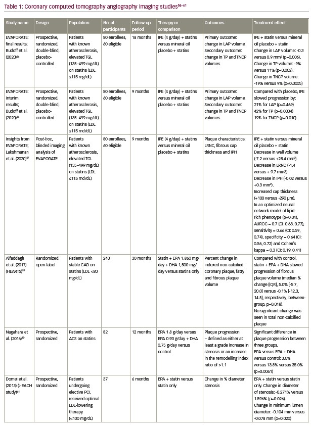
The EVAPORATE (Effect of icosapent ethyl on progression of coronary atherosclerosis in patients with elevated triglycerides on statin therapy) trial compared the effect of high-dose (4 g/day) pure IPE versus placebo (mineral oil) on plaque characteristics using serial coronary computed tomography angiography (CCTA).56 Patients were previously on statin therapy, had known atherosclerotic CAD and had triglyceride levels 135–499 mg/dL and LDL levels ≤115 mg/dL. Compared with placebo, IPE 4 g/day reduced low attenuation plaque volume (-0.3 mm3 versus 0.9 mm3, respectively; p=0.006) at 18 months, as measured by CCTA. Total plaque volume and total non-calcified plaque volume also reduced. Furthermore, a novel software (Elucid Bioimaging Inc., Boston, MA, USA) validated using histology, was used to evaluate the vulnerable plaque features in the EVAPORATE trial. Post hoc imaging analysis revealed decreased lipid-rich necrotic core (-1.4 mm3 versus 9.7 mm3) and intraplaque haemorrhage (-0.02 mm3 versus 0.3 mm3), and increased fibrous cap thickness (100 versus -290 µm) between the treatment and placebo groups, respectively; all these characteristics confer greater plaque stability.57
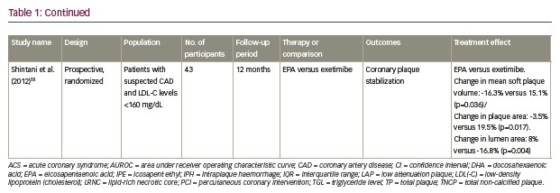
Similarly, Shintani et al. performed another prospective randomized study, which enrolled patients with suspected CAD and an LDL cholesterol level <160 mg/dL.58 Patients were randomized to receive EPA or ezetimibe. Coronary plaque was evaluated by multidetector computed tomography at baseline and 1 year follow-up. This study demonstrated a significant improvement in plaque area (percent change -3.5% versus 19.5%; p=0.017), lumen area (percent change 8% versus -16%; p=0.004) and soft-plaque volume (percent change -16.3% versus 15.1%; p=0.036) in the EPA group compared with the ezetimibe group, respectively.58 Both the EVAPORATE and the Shintani et al. studies showed that EPA conferred greater plaque stability than either placebo or ezetimibe. One difference between these two trials is that the EVAPORATE trial used the pure form of IPE. Additionally, the Shintani et al. study suggested that the beneficial effects on coronary plaque stabilization are independent from LDL cholesterol level, and EPA lowered the incidence of major cardiovascular events compared with the ezetimibe (control) group.58
Alfaddagh et al. investigated whether adding omega-3 ethyl ester to statin therapy in patients with CAD slowed the progression of coronary plaque volume.59 In the intention-to-treat analysis, there was no difference between the OM3FAs plus statin and the statin-only group (p=0.14) in the primary outcome of non-calcified plaque volume but these findings approached significance in the per-protocol analysis (p=0.07). Additional analysis was done to see whether there were differences in the formation of fatty and fibrous plaque as measured by CCTA. Fibrous plaque progression was more pronounced in the control group than in those receiving omega-3 ethyl ester (p=0.018). In this study, high-dose EPA + DHA (3.36 g/day) provided additional benefit to statins in preventing the progression of fibrous coronary plaque over 30 months in patients with CAD and LDL cholesterol <80 mg/dL. It is believed that resolvins synthesized from EPA and DHA accelerate the resolution of inflammation, which may reduce fibrosis. Interestingly, this effect was observed only in individuals taking low-intensity statins; those receiving high-intensity statins were unaffected. These findings imply that the interplay between statin treatment and OM3FAs may play a significant role in determining plaque volume and clinical outcomes. This study was limited by its open-label design, which may have influenced the results by allowing controls to opt in to treatment.59
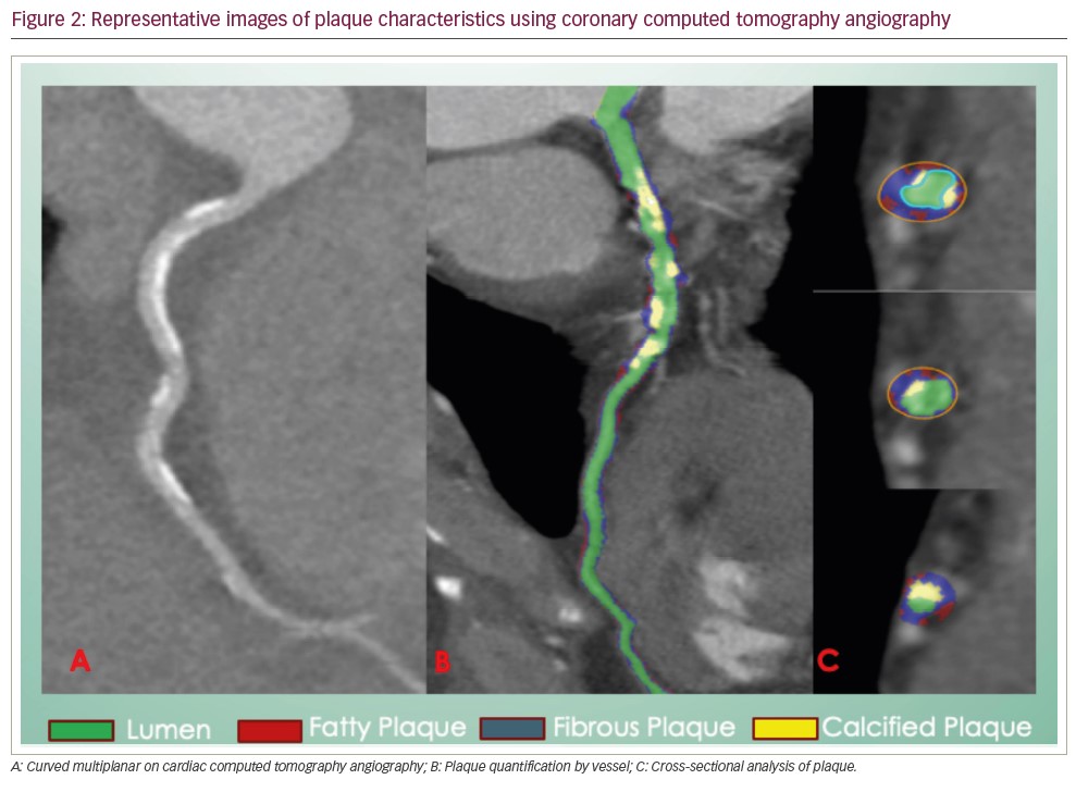
Nagahara et al. studied patients with acute coronary syndrome on statin therapy using CCTA (Figure 2).60 Patients were divided into groups comparing EPA 1.8 g/day, EPA 0.93 g/day + DHA 0.75 g/day and control (statin only), and followed for 12 months. Plaque progression was defined as at least a grade increase in stenosis or an increase of >1.1 in the remodelling index ratio. There was a significant difference in progression of plaque between the three groups, with the lowest being 3% in the EPA group followed by 13% in the EPA + DHA group and 35% in the control group (p=0.0061). These results emphasize the distinct effects of EPA + DHA versus EPA alone.60
Similarly, Domei et al. evaluated the effects of EPA and statin versus statin-only therapy in patients who underwent elective percutaneous coronary intervention (PCI).61 There was no difference in LDL cholesterol level between the two groups at the beginning of the study. However, a significant difference in minimum luminal diameter and percent diameter stenosis using quantitative coronary analysis of CCTA was noted at the 6-month follow-up. The change in lumen diameter was -0.104 mm versus -0.078 mm (p=0.020) and the change in percent diameter stenosis was -0.271% versus 1.596% between the treatment and control groups, respectively.61
In the AQUAMARINE EPA/DHA (Effect of eicosapentaenoic acid/docosahexaenoic acid on coronary high-intensity plaques detected with non-contrast T1-weighted imaging) study, a recent single-centre randomized controlled trial, the effect of EPA + DHA on high-risk coronary plaques was investigated using magnetic resonance angiography and CCTA in patients with CAD on statin therapy.62 Patients were randomized to 2 g/day EPA + DHA, 4 g/day EPA + DHA or placebo. In CCTA plaque analysis, there was no significant change detected in total plaque volume, low attenuation plaque volume, vessel area or plaque area from baseline to 12 months in each group. These findings are likely related to the eligibility criteria of the study where participants were not limited to high triglyceride levels. Additionally, distinct physiological effects of EPA and DHA may have contributed to the reported results.62
The main advantages of CCTA include its non-invasive and inexpensive nature and the ease with which it is performed. Additionally, intracoronary evaluation of multiple arteries with intravascular ultrasound (IVUS) and intravascular optical coherence tomography (OCT) is inconvenient and not clinically indicated in all patients having cardiac catheterization. Moreover, serial imaging using CCTA is simple and safe, and can be used to evaluate vulnerable lesions and track the effects of therapy (Table 1).63
Intravascular ultrasound and integrated backscatter intravascular ultrasound
IVUS and integrated backscatter IVUS (IB-IVUS) imaging techniques can also assess atherosclerotic plaque characteristics. IVUS is a miniature ultrasound device with a transducer attached to the end of the catheter, allowing for imaging of the coronary artery wall and measuring wall thickness.64 IB-IVUS provides colour-coded images showing plaque composition, intimal hyperplasia, lipid core, fibrous cap, mixed lesions and calcifications.65
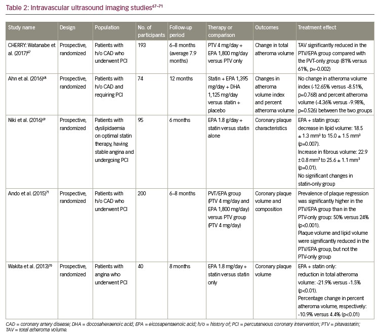
IVUS investigations have shown that high-dose statin treatment stabilizes and regresses coronary artery plaque.66 IB-IVUS was used in the CHERRY (Combination therapy of eicosapentaenoic acid and pitavastatin for coronary plaque regression evaluated by integrated backscatter intravascular ultrasonography) study to determine whether EPA supplementation added to high-dose pitavastatin therapy enhances coronary plaque regression and stabilization compared with high-dose pitavastatin alone.67 Participants had known CAD and PCI. There was a 6- to 8-month follow-up period, with an average follow-up of 7.9 months. The normalized total atheroma volume (TAV) was significantly lower in both groups, according to the volumetric IVUS analysis. In addition, TAV regression was also more prevalent in the EPA and pitavastatin group than in the pitavastatin group (81% versus 61%, respectively), and this difference was statistically significant (p=0.002).67
Another trial used IB-IVUS to track plaque progression in patients with known CAD who needed PCI. All patients were prescribed statin and randomly assigned to either the n-3 group, which received 3 g/day of omega-3 PUFA comprising 1,395 mg EPA and 1,125 mg DHA, or the placebo group. Patients were monitored for 12 months. Adding OM3FAs to statin therapy did not affect atheroma volume index or change in percent atheroma volume.68 These findings are possibly explained by varying amounts of fish in patients’ diets, particularly fish high in OM3FAs. For example, in Korea, the omega-3 index is high because of the traditionally fish-heavy diet. Patients in the placebo group had greater levels of OM3FAs at baseline, possibly because of a diet heavy in fish, which may have prevented the combined therapy with EPA and statin from inducing synergistic effects.
Two studies by Niki et al.69 and Wakita et al.70 evaluated the effect of high-purity EPA (1.8 g/day) added to high-dose statin therapy on plaque characteristics using IB-IVUS/IVUS in patients with stable angina who underwent PCI. In Niki et al., patients were followed for 6 months and the patients’ coronary plaque characteristics were analysed using IB-IVUS before and after treatment.69 A significant reduction in lipid volume (18.5 ± 1.3 mm3 to 15.0 ± 1.5 mm3; p=0.007) and a significant increase in fibrous volume (22.9 ± 0.8 mm3 to 25.6 ± 1.1 mm3; p=0.01) were observed in the EPA group. No significant changes were seen in plaque components in the control group.69 Wakita et al. evaluated plaque regression using IVUS over 8 months.70 The EPA group received an additional 1,800 mg/day of EPA for more than 6 months compared with the statin-only control group. In all patients, IVUS was performed on the day of PCI and at an average of 8 months later. Coronary plaque volume significantly reduced in the EPA group compared with the non-EPA group (-21.9% versus -1.5% for TAV; -10.9% versus 4.4% for percent change in percent atheroma volume; p<0.01).70
Another large study evaluated the effect of high-dose pitavastatin (4 mg/day) and EPA (1.8 g/day) on coronary plaque regression using IB-IVUS.71 This multicentre study enrolled 200 patients who underwent PCI across six different hospitals and followed them for 8 months. Plaque volume and lipid volume were significantly reduced in the pitavastatin/EPA group but not in the pitavastatin-only group. Plaque regression was defined as percent change in plaque volume more than -14.6%. The prevalence of plaque regression was significantly higher in the pitavastatin/EPA group than in the pitavastatin group (50% versus 24%, respectively; p<0.001). A multivariate regression analysis demonstrated that adding EPA was independently associated with plaque regression after adjusting for confounding factors.71
The primary beneficial characteristics of IVUS are its ability to quantify the percentage of narrowing and to give insight into the nature of the plaque. However, it is limited by its invasive nature, operator expertise and measurement variability during systole and diastole of the cardiac cycle (Table 2).65
Optical coherence tomography
OCT is an imaging technique that uses emission and reflection of near-infrared light to produce cross-sectional images of coronary arteries to evaluate plaque characteristics.72 OCT has approximately 10-fold greater resolution than ultrasound-based techniques, with a resolution of 4–20 μm. This makes it an ideal imaging modality to identify the thin fibrous cap (defined as <65 μm) in patients with thin-cap fibroatheroma (Table 3).73
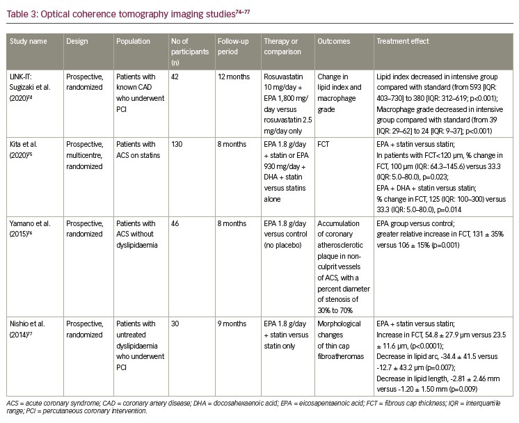
A subgroup analysis of 42 patients from the LINK-IT (Effect of rosuvastatin and eicosapentaenoic acid on neoatherosclerosis) trial compared changes in the lipid index and macrophage grade of native coronary plaques detected by OCT in those receiving intensive therapy (rosuvastatin plus EPA) and those receiving standard therapy (rosuvastatin alone) over a 12 month period.74 Between baseline and follow-up, the lipid index decreased considerably in the intensive-therapy group (from 593 [interquartile range (IQR) 403–730] to 380 [IQR 312–619]; p=0.001), but not in the standard-therapy group (from 660 [IQR 219–825] to 601 [IQR 242–898]; p=0.078). Macrophage grade reduced significantly between baseline and follow-up in the intensive-therapy group (from 39 [IQR 29–62] to 24 [IQR 9–37]; p=0.001) but remained stable in the statin-therapy group (from 44 [IQR 30–58] to 51 [IQR 21–66]; p=0.364). This study also found that changes in lipid index and macrophage grade in native coronary plaques associated positively with those in neoatherosclerotic plaques.74
Kita et al. enrolled 130 patients with acute coronary syndrome who were treated with statins.75 Patients were assigned to a statin-only group, statin plus high-dose EPA (1,800 mg/day) or statin plus EPA (930 mg/day) plus DHA (750 mg/day). OCT was performed at baseline and at 8 months’ follow-up. There was no significant difference in percent change of minimum fibrous cap thickness between the groups. However, the percent change for minimum fibrous cap thickness was significantly higher in the EPA group with thinner fibrous caps (<120 μm). EPA plus statin versus statin alone showed a percent change in fibrous cap thickness of 110% (IQR 64–145%) versus 33% (IQR 5–80%; p=0.0230). EPA plus DHA plus statin versus statin alone showed a percent change in fibrous cap thickness of 125% (IQR 100–300%) versus 33% (IQR 5–80%; p=0.014).75 In contrast, Yamano et al. also enrolled patients with acute coronary syndrome but without dyslipidaemia.76 Patients were divided into two groups: EPA (1,800 mg/day) and a non-EPA control group. OCT was performed at baseline and at 8 months’ follow-up. Fibrous cap thickness significantly increased in both the EPA and control groups. However, the relative change in fibrous cap thickness was significantly greater in the EPA group than in the control group (131 ± 35% versus 106 ± 15%; p=0.001).76
Another study evaluated the effect of combined EPA and statin therapy on thin fibrous cap atheroma in patients with untreated dyslipidaemia who underwent PCI.77 Patients were randomly assigned to EPA (1,800 mg/day) plus statin or statin alone and followed for 9 months. OCT analysis showed that, compared with the statin-only group, the EPA plus statin group had a greater increase in fibrous cap thickness (54.8 ± 27.9 μm versus 23.5 ± 11.6 μm; p<0.0001), with a greater decrease in lipid arc (-34.4 ± 41.5 μm versus -12.7 ± 43.2 μm; p=0.007) and lipid length (2.81 ± 2.46 mm versus -1.20 ± 1.50 mm; p=0.009).77 Similarly, Uehara et al. enrolled patients with stable angina on statin therapy.78 Patients were randomly assigned to EPA plus statin or a statin-only group, and morphological changes of thin fibrous cap atheroma were measured. Both groups had significant fibrous cap thickness; however, the difference in the change of fibrous cap thickness was highly significant in the EPA plus statin group versus the statin-only group (106 ± 19 μm versus 26 ± 9 μm, respectively; p=0.0003). The change in the lipid arc also decreased significantly in the EPA plus statin group compared with the control group (-40.0 ± 11.5 versus -4.0 ± 5.5; p=0.0074).78
Carotid ultrasound
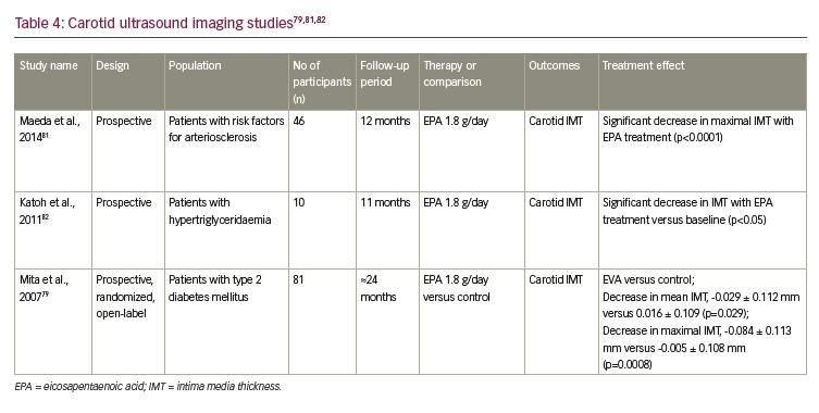
The effect of EPA on atherosclerosis was also evaluated in a selective group of patients by measuring carotid intima media thickness (IMT) with carotid ultrasound.79,80 IMT is the thickness of the tunica intima and tunica media, the innermost two layers of artery walls. Carotid IMT is used to detect atherosclerosis. A prospective trial evaluated patients with atherosclerotic risk factors who received treatment with EPA (1.8 g/day) and were followed up for 12 months. There was significant decrease in maximal IMT from baseline with EPA treatment (p<0.0001).81 Similarly, two other randomized trials, Katoh et al.82 and Mita et al.,79 evaluated patients with hypertriglyceridaemia (11-month follow-up) and type 2 diabetes (24-month follow-up), respectively, with the treatment group receiving 1.8 g/day of EPA versus control. In Katoh et al., there was a significant decrease in IMT in the treatment group compared with baseline (p<0.05).82 In Mita et al., there was significant decrease in mean IMT (-0.029 ± 0.112 mm versus 0.016 ± 0.109 mm; p=0.029), as well as a significant decrease in maximum IMT (-0.084 ± 0.113 mm versus -0.005 ± 0.108 mm; p=0.0008) compared with the control group.79 Although carotid IMT predicts future cardiovascular events, the precise atherosclerotic characteristics may differ in carotid arteries compared with coronary arteries. In addition, carotid IMT lacks sensitivity and specificity in detecting atherosclerosis.83 The relationship between carotid and coronary artery atherosclerosis and their role in altering cardiovascular events needs to be explored further. Despite this, EPA seems to be beneficial in improving carotid IMT, which is a known surrogate for atherosclerosis (Table 4).84
Cardiac magnetic resonance
Cardiac magnetic resonance can identify high-risk coronary plaques. These high-risk/high-intensity plaques are quantitatively assessed using the plaque-to-myocardium signal-intensity ratio (PMR). These high-intensity plaques with PMR ≥1.4 are associated with future coronary events.85 The anti-atherosclerotic impact of high-dose EPA/DHA in patients with CAD who were receiving statin therapy was assessed in a post hoc subgroup analysis of the AQUAMARINE EPA/DHA study in patients with elevated triglycerides (>150 mg/dL).86 Participants were randomly assigned to receive either 2 g/day of EPA/DHA (n=26), 4 g/day of EPA/DHA (n=23) or statin only (n=24). At 12 months, high-dose EPA/DHA therapy significantly reduced PMR compared with the statin-only control. This study supports the role of higher doses of EPA/DHA in lowering residual risk in patients with CAD on statin therapy.86
Conclusion
As research into the optimal management of dyslipidaemia develops, there is a need for additional therapy beyond statins to reduce the risk of atherosclerotic CVD. In large cardiovascular outcome trials, EPA has demonstrated cardiovascular benefit.49,50 Furthermore, multiple serial-imaging studies have shown benefits of OM3FAs on plaque progression and characteristics. IPE especially has favourable efficacy and safety, and is superior to other OM3FAs in reducing cardiovascular risk. Future research should compare EPA with EPA + DHA to account for the contradictory outcomes observed in some of the outcome trials with EPA + DHA.52 Additionally, we must acknowledge that the pleiotropic effects of IPE may have contributed to the disparities between trials. Current research and accumulating evidence support IPE as an adjunct therapy to statins to reduce residual cardiovascular risk. ❑


