touchCARDIO touchCARDIO
Arrhythmia
Read Time: 2 mins

73/Automated, high-precision echocardiographic and haemodynamic atrioventricular delay assessment for right ventricular pacing in hypertrophic obstructive cardiomyopathy

Copy Link
Published Online: Oct 9th 2012 European Journal of Arrhythmia & Electrophysiology. 2022;8(Suppl. 1):abstr73
Authors: JS Mohal (Presenting Author) – National Heart & Lung Institute, Imperial College London, London; F Simader – National Heart & Lung Institute, Imperial College London, London; M Shun-Shin – National Heart & Lung Institute, Imperial College London, London; A Arnold – National Heart & Lung Institute, Imperial College London, London; Z Whinnett – National Heart & Lung Institute, Imperial College London, London
Quick Links:
Article
Article Information
Article:

Background: Right ventricular pacing (RVP) to reduce left ventricular outflow tract (LVOT) obstruction in hypertrophic obstructed cardiomyopathy (HOCM) remains controversial. Imprecise and variable atrioventricular delay (AVD) selection methods have contributed to the uncertainty. Using automated, high-precision AVD selection methods that target cardiac output incrementation may allow reliable identification of the optimal AVD to determine whether optimised RVP is beneficial in HOCM.

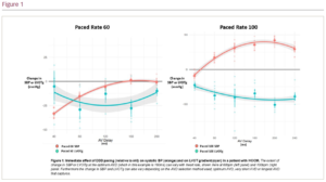
Methods: A total of 15 patients with HOCM and dual chamber pacing devices in situ were recruited. Pacing was alternated between atrial pacing (AAI) and AV sequential RVP (DDD) 10 times whilst beat-by-beat blood pressure (BP) via non-invasive finometry and LVOT continuous wave Doppler for LVOT gradient (LVOTg) via echocardiography were continuously recorded. This was repeated for a full range of AVDs at 10 bpm above sinus rate and 100 bpm. Custom software automated detection of LVOTg trace troughs and BP trace peaks to identify beat-by-beat LVOTg and systolic blood pressure (SBP), respectively. The mean difference in LVOTg and BP between pacing states (AAI and DDD) were plotted for each AVD and fitted to quadratic curves to improve precision (Figure 1). The peak of SBP curve identified the optimal AVD for cardiac output. Averaging changes in beat-by-beat SBP and LVOTg across multiple pacing transitions accounts for physiological drift and automated analysis reduces bias.

Results: Mean LVOTg of recruited patients was 50 mmHg at rest. At 10 bpm above sinus rate, DDD pacing at optimal AVD significantly increased SBP (+2.72 ± 1.33 mmHg; p=0.05) and significantly reduced LVOTg (-12.4 ± 3.64 mmHg; p=0.002). At 100 bpm, greater SBP increases were seen (+10.0 ± 3.96 mmHg; p=0.02) with greater LVOTg reductions (-23.7 ± 6.95 mmHg; p=0.004). Alternative described methods for AVD selection also reduced LVOTg but with worse SBP responses. Longest fully captured AVD reduced LVOTg (-3.34 ± 2.05 mmHg) but increased SBP by a smaller amount than optimal AVD (+1.36 ± 1.1 mmHg). Very short AVD (40–80 ms) reduced LVOTg (-5.55 ± 4.3 mmHg; p=0.2) but substantially reduced SBP (-23.5 ± 3.22 mmHg; p<0.01).

Conclusion: High precision, automated haemodynamic measurements detected the optimum AV delay that balanced the increased LVOT size, reduced myocardial performance and altered AV filling that occur with RVP for HOCM. AVD-optimised RVP reduced LVOTg and improved cardiac output. 

Further Resources

Share this Article
Related Content In Arrhythmia
  • Copied to clipboard!
    accredited arrow-down-editablearrow-downarrow_leftarrow-right-bluearrow-right-dark-bluearrow-right-greenarrow-right-greyarrow-right-orangearrow-right-whitearrow-right-bluearrow-up-orangeavatarcalendarchevron-down consultant-pathologist-nurseconsultant-pathologistcrosscrossdownloademailexclaimationfeedbackfiltergraph-arrowinterviewslinkmdt_iconmenumore_dots nurse-consultantpadlock patient-advocate-pathologistpatient-consultantpatientperson pharmacist-nurseplay_buttonplay-colour-tmcplay-colourAsset 1podcastprinter scenerysearch share single-doctor social_facebooksocial_googleplussocial_instagramsocial_linkedin_altsocial_linkedin_altsocial_pinterestlogo-twitter-glyph-32social_youtubeshape-star (1)tick-bluetick-orangetick-red tick-whiteticktimetranscriptup-arrowwebinar Sponsored Department Location NEW TMM Corporate Services Icons-07NEW TMM Corporate Services Icons-08NEW TMM Corporate Services Icons-09NEW TMM Corporate Services Icons-10NEW TMM Corporate Services Icons-11NEW TMM Corporate Services Icons-12Salary £ TMM-Corp-Site-Icons-01TMM-Corp-Site-Icons-02TMM-Corp-Site-Icons-03TMM-Corp-Site-Icons-04TMM-Corp-Site-Icons-05TMM-Corp-Site-Icons-06TMM-Corp-Site-Icons-07TMM-Corp-Site-Icons-08TMM-Corp-Site-Icons-09TMM-Corp-Site-Icons-10TMM-Corp-Site-Icons-11TMM-Corp-Site-Icons-12TMM-Corp-Site-Icons-13TMM-Corp-Site-Icons-14TMM-Corp-Site-Icons-15TMM-Corp-Site-Icons-16TMM-Corp-Site-Icons-17TMM-Corp-Site-Icons-18TMM-Corp-Site-Icons-19TMM-Corp-Site-Icons-20TMM-Corp-Site-Icons-21TMM-Corp-Site-Icons-22TMM-Corp-Site-Icons-23TMM-Corp-Site-Icons-24TMM-Corp-Site-Icons-25TMM-Corp-Site-Icons-26TMM-Corp-Site-Icons-27TMM-Corp-Site-Icons-28TMM-Corp-Site-Icons-29TMM-Corp-Site-Icons-30TMM-Corp-Site-Icons-31TMM-Corp-Site-Icons-32TMM-Corp-Site-Icons-33TMM-Corp-Site-Icons-34TMM-Corp-Site-Icons-35TMM-Corp-Site-Icons-36TMM-Corp-Site-Icons-37TMM-Corp-Site-Icons-38TMM-Corp-Site-Icons-39TMM-Corp-Site-Icons-40TMM-Corp-Site-Icons-41TMM-Corp-Site-Icons-42TMM-Corp-Site-Icons-43TMM-Corp-Site-Icons-44TMM-Corp-Site-Icons-45TMM-Corp-Site-Icons-46TMM-Corp-Site-Icons-47TMM-Corp-Site-Icons-48TMM-Corp-Site-Icons-49TMM-Corp-Site-Icons-50TMM-Corp-Site-Icons-51TMM-Corp-Site-Icons-52TMM-Corp-Site-Icons-53TMM-Corp-Site-Icons-54TMM-Corp-Site-Icons-55TMM-Corp-Site-Icons-56TMM-Corp-Site-Icons-57TMM-Corp-Site-Icons-58TMM-Corp-Site-Icons-59TMM-Corp-Site-Icons-60TMM-Corp-Site-Icons-61TMM-Corp-Site-Icons-62TMM-Corp-Site-Icons-63TMM-Corp-Site-Icons-64TMM-Corp-Site-Icons-65TMM-Corp-Site-Icons-66TMM-Corp-Site-Icons-67TMM-Corp-Site-Icons-68TMM-Corp-Site-Icons-69TMM-Corp-Site-Icons-70TMM-Corp-Site-Icons-71TMM-Corp-Site-Icons-72