Background: Obesity has been strongly associated with atrial and ventricular arrhythmias. This effect is, in part, mediated by epicardial adipose tissue (EAT), which has previously shown to have a pro-arrhythmic secretome. Therefore, we sought to confirm any paracrine effects of adipose tissue on myocardial conduction and tested the differential effects of human EAT and subcutaneous adipose tissue (SAT) on ventricular myocyte electrophysiology.
Methods: EAT and SAT harvested from 11 patients during cardiothoracic surgery were cultured to generate adipose tissue-conditioned media (Cme), and their secretomes characterised using adipokine arrays and ELISA. Conduction velocities in monolayers of neonatal rat ventricular myocytes (NRVM) were measured at baseline and at 2, 4 and 24 hours post-incubation with EAT-, SAT- and control-CMe.
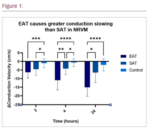
Results: Referenced to baseline at 2 hours, NRVM exposed to EAT- and SAT-Cme recorded slower conduction velocities than control, though they were comparable decrements (-6.3 ± 3.4 cm/s and -4.5 ± 3.6 cm/s vs -1.0 ± 2.7 cm/s; p=0.0001 and p=0.014, respectively). At 4 hours, NRVM incubated with EAT-Cme demonstrated greater conduction slowing than with SAT-Cme (-10.9 ± 5.7 cm/s vs -4.2 ± 3.4 cm/s; p=0.0024). At 24 hours, further reduction in conduction velocity was observed, with EAT again inducing a greater effect than SAT (-15.1 ± 5.2 cm/s vs -6.4 ± 5.8 cm/s;
p=0.011). Electrogram amplitude, duration and fractionation exhibited no significant difference between all groups. There were no significant differences in the adipokine secretomes of EAT and SAT. Moreover, stratification of the study cohort by mean age and BMI, and the presence of cardiometabolic disease, demonstrated comparable adipokine profiles of EAT and SAT, respectively.
Conclusion: Acute exposure to adipose-Cme causes conduction slowing in vitro, which is more marked with EAT- vs SAT-Cme. These results confirm the arrhythmogenic effect of EAT, which is mediated, in part, by a paracrine mechanism. Future studies are required to determine the mediators of the paracrine effect and investigate potential therapeutic targets.